Xiang-Dong Guo1 ![]() ,
Qing-Lin Wang2,
Ying Li1,
Zhi-Cheng Zhang1
,
Qing-Lin Wang2,
Ying Li1,
Zhi-Cheng Zhang1
For correspondence:- Xiang-Dong Guo Email: gxdguoxd@163.com
Received: 12 December 2015 Accepted: 12 July 2016 Published: 30 August 2016
Citation: Guo X, Wang Q, Li Y, Zhang Z. Clinical effect of prootic and opisthotic injection of dexamethasone and gentamicin in meniere disease therapy. Trop J Pharm Res 2016; 15(8):1781-1786 doi: 10.4314/tjpr.v15i8.26
© 2016 The authors.
This is an Open Access article that uses a funding model which does not charge readers or their institutions for access and distributed under the terms of the Creative Commons Attribution License (http://creativecommons.org/licenses/by/4.0) and the Budapest Open Access Initiative (http://www.budapestopenaccessinitiative.org/read), which permit unrestricted use, distribution, and reproduction in any medium, provided the original work is properly credited..
Purpose: To evaluate the effect of prootic and opisthotic injection of dexamethasone and gentamicin for the treatment of Meniere disease.
Methods: A total of 192 cases of Meniere disease were randomly selected from The First Affiliated Hospital of Henan University of TCM between April 2012 and December 2014. They were divided into three groups, i.e., dexamethasone (DX group), gentamicin (GM group) and normal saline (NS group). According to the test design, patients in the DX and GM groups were administered prootic and opisthotic injection of dexamethasone and gentamicin (5.5 mL once a week), respectively, for four weeks. Changes in auditory and vestibular functions, as well as control of capacity for action, were observed.
Results: On the 18th day after treatment, dizziness control rates in the DX group, GM group, and NS group were 72.73, 75.86 and 12.12 %, respectively; differences between the DX group and NS group and between the GM group and NS group were significant (p < 0.01). DX and GM groups both showed effective control of dizziness. Hearing improvement rate in DX, GM and NS groups was 39.39, 6.89 and 9.09 %, respectively (p < 0.01), indicating that only DX group showed effective hearing improvement. Improvement rate of capacity for action in DX, GM and NS groups was 75.76, 72.41 and 15.15 %, respectively; differences in the control rate of capacity for action between DX and GM groups and between GM and NS groups were significantly different (p < 0.01). Thus, DX and GM groups showed effective control of the capacity for action.
Conclusion: Prootic and opisthotic injection of dexamethasone and gentamicin is a potentially safe and effective new approach for the treatment of Meniere disease.
Introduction
Meniere disease is an inner ear disease characterized by idiopathic endolymphatic hydrops with symptoms such as recurrent rotatory vertigo, fluctuating sensorineural hearing loss, tinnitus, and/or sensation of ear fullness [1]. Although it is known to be induced by endolymphatic hydrops, the pathogenesis has not been thoroughly studied and its development is unpredictable. In recent years, Meniere disease has been thought to be caused by endolymphatic hydrops resulting from immune complex deposition in the stria vascularis and endolymphatic sac, which is the result of autoimmunity or a subclinical type virus-induced immune reaction [2,3]. It is also believed that Meniere disease is associated with inflammation and allergic reaction. The treatment goal of Meniere disease is mainly to control dizziness and resolve residual hearing; however, as the pathogenesis of dizziness remains unclear, the processing method is clinically diverse [4]. Frequent attacks of Meniere disease can lead to permanent and irreversible hearing impairment that cannot be cured. Although Meniere disease cannot be cured, treatment can control or relieve symptoms [5]. Systematic medication and local medication are currently the major methods for treating Meniere disease.
In the last few decades, intratympanic injection has been shown to be effective in treating Meniere disease, but it may lead to complications such as tympanic membrane perforation, otitis media, labyrinthitis, and hearing loss; head position and auditory tube opening can influence the amount of drug that accesses the inner ear. Yang Xiaoqi [6] from China found that drug can fully enter the inner ear if being injected behind the ears. Over the last three years, our hospital has attempted to treat Meniere disease through prootic and opisthotic injection of dexamethasone and gentamicin, and has obtained satisfactory results.
Methods
General data
A total of 192 patients with Meniere disease who received treatment at department of otorhinolaryngology of The First Affiliated Hospital of Henan University of TCM from April 2012 to December 2014 were selected as research subjects [7]. All patients were confirmed to have Meniere disease based on the Diagnostics Basis and Curative Effect Appraisal of Meniere Disease formulated by the Guiyang Conference in 2006. Moreover, they must have had the disease for at least 0.5 years and received drug treatment for more than 6 months. Cases with dizziness due to other reasons were excluded. All patients signed an informed consent form. Before treatment, all patients underwent pure tone audiometry and auditory brainstem response tests, and were shown to have an auditory threshold of affected ear over 40 dbHL and healthy ear below 30 dBHL. All conventional drug treatments were stopped during treatment. Patients were divided into the dexamethasone (DX group), gentamicin (GM group), and normal saline (NS group). In the DX group (66 cases), 34 cases were male and 32 were female, with ages ranging from 26 to 61 years (mean (41.3 ± 0.7) years) and disease course ranging from 1 to 11 years. In the GM group (60 cases), 26 cases were male and 34 were female, with age ranging from 29 to 63 years (mean (43.6 ± 0.9) years) and disease course ranging from 1 to 13 years. In the NS group (66 cases), 30 cases were male and 36 cases were female, with age ranging from 27 to 62 years (mean (41.9 ± 1.1) years) and disease course ranging from 1 to 12 years. Differences in the number of cases, body mass, gender, and age between the three groups showed no statistical significance (p > 0.05); therefore, the results were comparable. The study was approved by the Ethics Committee of the First Affiliated Hospital of Henan University of TCM (Approval No.: 20150221GXD) and it followed the Declaration of Helsinki [8].
Method
Drug delivery in all groups followed specific stipulations for the injection site and method. For prootic injection, drugs were injected through the site ahead of the superior notch of the tragus and the depression above the mandibular condyle. With regard to opisthotic injection, drugs were injected through the lower part, ahead of the mastoid process behind the ears and depression at the inferior margin behind the earlobe. The respective drugs were first absorbed into a sterile syringe equipped with No. 5 skin-test needle and then injected through the sites mentioned above. The needle was vertically inserted into the skin for 0.5 cm. The drug was injected if there was no blood flowing back (0.5 mL each side). Intensive twirling, lifting, and thrusting was not allowed. The injection was performed once a week for four weeks. Patients in the DX group were administered 1 mL dexamethasone (10 mg/mL) and the GM group was administered 1 mL gentamicin (30 mg/mL) consisting of 1 mL gentamicin (40 mg/mL) and 0.5 mL sodium bicarbonate buffer (0.6 mol/L). The NS group was administered 1 mL normal saline (0.9 %). Injection was stopped if the following were observed: positive results based on the spontaneous nystagmus and head-shaking test, obvious decline in hearing, and significantly improved subjective symptoms. Two patients in the GM group stopped treatment as they experienced significantly decreased hearing after the second injection.
Evaluation of curative effect
Curative effect was evaluated according to the Diagnostic Basis and Curative Effect Appraisal of Meniere Disease formulated by the Guiyang Conference in 2006. Onset and frequency of dizziness 18 - 24 months after treatment as well as six months before treatment were compared. Degree of dizziness could be divided into five levels: completely controlled, basically controlled, partially controlled, uncontrolled, and worse. Hearing was evaluated based on results obtained by the average hearing threshold value at 0.25, 0.5, 1, 2, and 3 kHz minus the poorest average hearing threshold 18 - 24 months after treatment.
Hearing improvement was divided into four levels: level A: improved more than 30 dB or hearing threshold lower than 20 dB at all frequencies; level B: improved 15 - 30 dB; level C: improved 0 - 14 dB (ineffective); level D: improved 0 dB (worse). Capacity of action was evaluated by comparing activity limited days from 18 to 24 months after treatment and activity limited days six months before treatment. Capacity for action could be divided into five levels; completely improved, basically improved, partially improved, unimproved, and worse.
Statistical analysis
SPSS19.0 software was used to process data. Enumerated data were expressed as percentages. Comparison between groups was performed using the χ2 test. When n < 5, the Fisher test was used. Differences were considered significant if p < 0.05.
Results
Dizziness
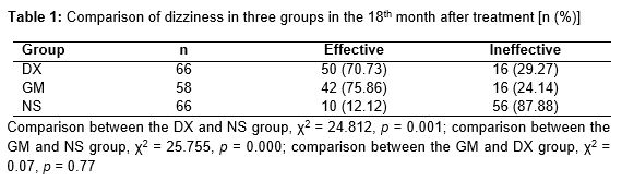
Of 192 cases, all patients completed the treatment with the exception of two cases in the GM group who rejected the treatment due to significantly decreased hearing after the second gentamicin injection. Dizziness control rate in the DX group, GM group, and NS group were 72.73, 75.87, and 12.12 %, respectively. Differences between the DX group and NS group and between the GM group and NS group were significant (p < 0.01). The difference between the DX group and GM group showed no significant difference (p > 0.05). Excluding the NS group, the other two groups showed obvious improvements in dizziness control ().
Hearing improvement
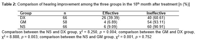
Pure tone audiometry (PTA) results of patients in the DX group were (46.67 ± 6.32) dBHL before treatment and (39.53 ± 8.57) dBHL in the 18th month after treatment; the GM group was (47.86 ± 6.97) dBHL and (46.56 ± 7.39) dBHL; and the NS group was (47.23 ± 7.11) dBHL and (48.52 ± 8.57) dBHL, respectively. In the 18th month after treatment, hearing improvement rates in the DX group, GM group, and NS group were 39.39, 6.89 and 9.09 % respectively. Differences in the DX group compared with the GM group and NS group were statistically significant (p < 0.01), suggesting that the DX group showed effective hearing improvement ().
Evaluation of capacity for action
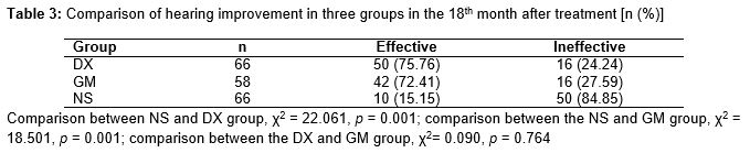
In the 18th month after treatment, action capacity improvement rates in the DX group, GM group, and NS group were 75.76, 72.41 and 15.15 %, respectively. There was a significant difference in action capacity control rates between the DX group and NS group and between the GM group and NS group (p < 0.01), suggesting that the DX group and GM group achieved effective control on action capacity ().
Incidence of untoward reactions
All patients in the GM group developed dizziness and gait instability within 2 days after the first injection, which was relieved after two weeks and totally recovered after three months. Moreover, two cases in the GM group showed significantly declined hearing after the second injection and stopped treatment. Patients in the DX group and NS group had no obvious unwanted reactions.
Discussion
Pathological changes in Meniere disease include endolymphatic hydrops. Currently, treatment methods for Meniere disease are diverse, including calcium antagonium and microcirculation improvement. However, all treatment methods can only control symptoms rather than cure the disease. Treatment with drugs is considered the major approach for treating Meniere disease, but the effects are not always curative [9,10]. Intratympanic injection has gradually become clinically important. In addition, the method has been supported based on sound effects [11].
Treating Meniere disease by injecting amino-glycosides through the tympanum is called chemical labyrinthectomy; injection of low dose gentamicin is effective in controlling symptoms and lowering the incidence of complications [12]. Besides puncture of tympanum, intratympanic injection can be performed through placement of a gelatin sponge containing medicinal preparation in a round window niche, tympanostomy tube insertion, or with a microperfusion pump. However, it is invasive and can easily cause infection and increase the incidence of thympanosclerosis. It has been reported that 40.4 % of patients who undergo repeated tympanotomy tube insertion develop tympanosclerosis [13]. Caye et al [14] noted that patients suffering from middle ear disease show improved hearing but a higher incidence of thympanosclerosis and atrophy after receiving tympanostomy tube insertion.
When patients with Meniere disease are treated with intratympanic administration [15], opening of the head and auditory tube will affect retention time and amount of drug in the tympanum, and permeability of the round window membrane will influence the amount of drug penetrating the inner ear. Yoshioka et al [16] found that permeability of the round window membrane differs among individuals. Among 55 patients (61 ears) with inner ear disease who were treated with intratympanic injection, 5 % of patients had no impermeable round window membrane and the round window membrane of 13% patients was poor. Nakashima et al [17] found that not all patients with inner ear disease were candidates for intratympanic administration.
Many studies have explored novel approaches for middle and inner ear administration to avoid complications. Recent results [18, 19] have shown that opisthotic injection of compound betamethasone shows an 82.5 % effectiveness rate in treating refractory low frequency sensorineural deafness. At the same drug dose, drug injected behind the ears more easily enters the sigmoid sinus, auditory vesicle, and perilymph fluid, and peaks earlier in local tissue and lasts longer compared to muscle injection. Opisthotic injection as a novel method for treating inner ear disease has shown good effects. In this study, we used prootic and opisthotic injection of dexamethasone and gentamicin to treat Meniere disease; dizziness control rate was 72.73 and 75.86 %, respectively, and improvement rate of capacity of action was 75.76 and 72.41 %, which are equivalent to results obtained in previous studies. After dexamethasone and gentamicin are injected around the ear, drugs enter the sigmoid sinus, tympanum, and inner ear and, thus, have an effect; in addition, there are typically no complications.
Limitations of the study
Steroid hormones, which act on the autoimmunity process and relieve abnormal immunity, cannot thoroughly change the physiopathologic mechanism and pathological progression of endolymphatic hydrops. Although the method can be used as a basic approach to treatment, prior to surgical operation for patients with refractory Meniere disease, the most effective dose and perfusion method should be further explored to achieve a long-lasting curative effect.
Conclusion
The findings of this study indicate that prootic and opisthotic injection of dexamethasone and gentamicin can effectively control dizziness, facilitate recovery of the physiological function of the inner ear, improve autonomic behavioral competence, and avoid risks and complications that may be induced by systematic administration and intratympanic administration. Therefore, it can be considered a simple, safe, and effective approach for managing Meniere disease.
Declarations
Acknowledgement
References
Archives


News Updates